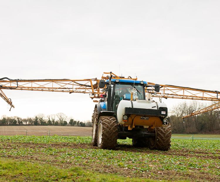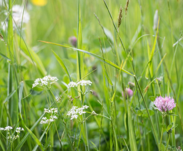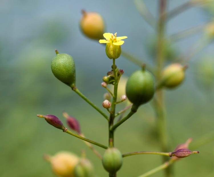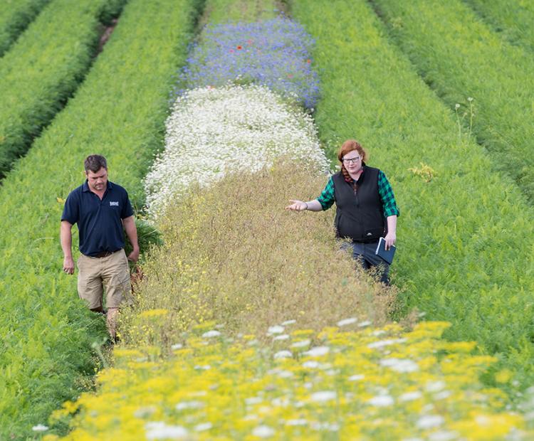
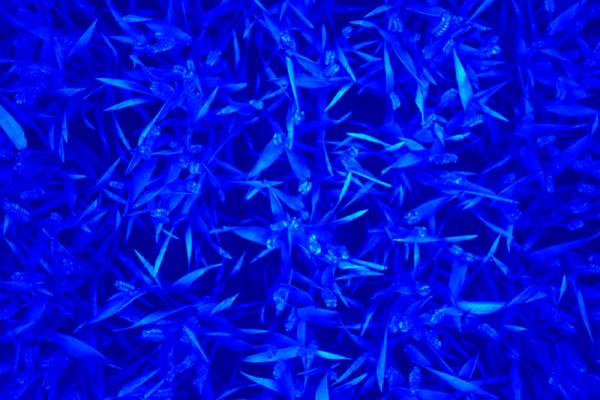
The big picture: using wildflower strips for pest control
They may be beautiful, but these strips prove pretty deadly for every farmer's mortal enemy: aphids
A high-resolution (3296 × 2472 px) 12 colour bit Prosilica GT3300 (Allied Vision, Stadtroda, Germany) with a maximum frame rate of 14.7 frames s-1 is employed as the visible camera. The camera is positioned perpendicular to the ground, and set up in auto-exposure mode to compensate outdoor light effects. Throughput: approximately 180 images h-1, inclusive of crane positioning time.
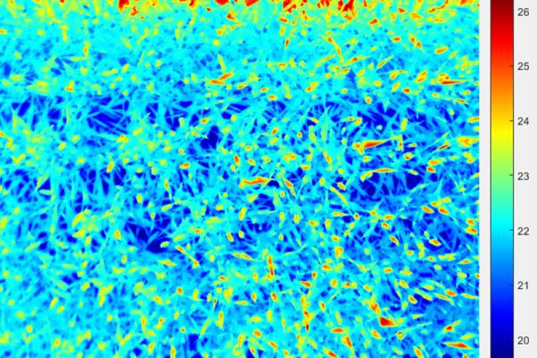
A FLIR A645SC (FLIR Systems Inc., Wilsonville, USA) is employed as a thermal infrared camera (640 × 480 px matrix) and covers the spectral range of 7.5 - 13 µm. The acquired thermal infrared images have a radiometric resolution of 0.05°C and an absolute precision of 2°C. Data is recorded in raw 16-bit format and the digital number intensities are later converted to radiometric temperature using the RBF equation provided by FLIR Systems. Throughput: approximately 330 images h-1, inclusive of crane positioning time.
Twin 3D laser scanners (Fraunhofer Institute, Munich, Germany) are mounted in the camera bay, opposing each other (see image below), and are capable of scanning plant canopies with high resolution (0.25 mm) in all three axes, using an NIR laser (840 nm) to ensure high reflectance by plant tissue and minimal physiological interaction. The laser scanners have a throughput of 30 plots h-1 and a field of view of approximately 0.5 m width and 0.5 m depth. Through the analysis of point cloud images, morphological traits of crops, such as plant height may be accurately quantified. Throughput: approximately 30 plots h-1, inclusive of crane positioning time.
The hyperspectral system is comprised of two mirror-scanning Hyperspec® Inspector (Headwall Photonic) Visible and near-infrared (VNIR) and Extended VNIR (ExVNIR) cameras, together covering the 400-1700 nm range. The VNIR and ExVNIR cameras have 1600/923 (0.7 nm step) and 320/229 (4.6 nm step) spatial/spectral resolution. During acquisition, the crane remains stationary and the resulting hypercube is collected from the motion of an internal concave mirror. Data is recorded in their original 16-bit format. Throughput: approximately 46 and 80 plots h-1 for the VNIR and ExVNIR, respectively, inclusive of crane repositioning time.
The on-board CFI (CropReporter™) is provided by PhenoVation (Wageningen, The Netherlands), and enables photosystem II (PSII) fluorescence measurements. The CFI is an LED (light emitting diode) induced chlorophyll fluorescent transient imager (Jalink and Schoor 2011) and chlorophyll fluorescence is induced by a flash of red light (620 nm) for 1400 ms, saturating the electron transfer between PSII and PSI (photosystem I) and records 24 images within that time. Throughput: approximately 90 plots h-1, inclusive of crane repositioning time.
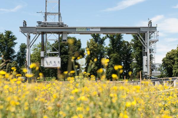
A four channel amplified radiometer light sensor (Skye Instruments Ltd., Powys, UK) is fitted on the Field Scanalyzer, with two channels calibrated for red (633 nm ± 19) wavebands and the others for NIR (800 nm ± 17), in order to automatically compute NDVI. One combination of waveband channels is positioned on top of the Field Scanalyzer, pointing at the sky to measure incident solar radiation, whilst the other pair points directly at the ground, to simultaneously measure radiation reflected upwards.