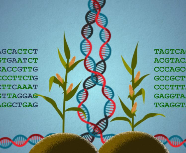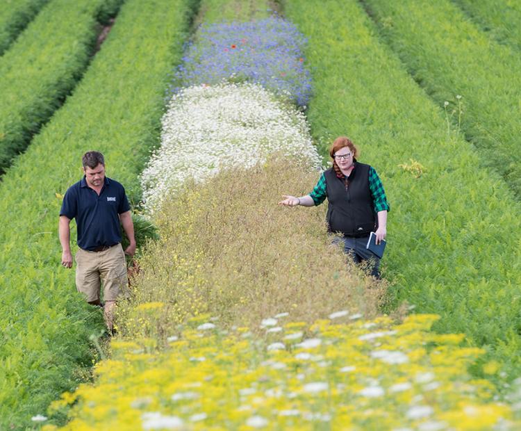
The big picture: using wildflower strips for pest control
They may be beautiful, but these strips prove pretty deadly for every farmer's mortal enemy: aphids
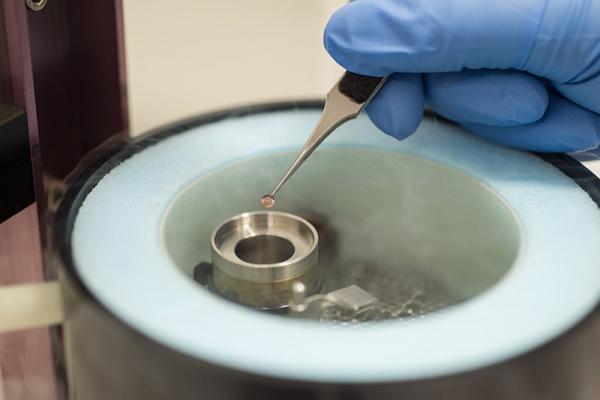
The Gatan Cryo Plunge can be used prepare frozen hydrated specimens for cryo TEM. Samples are loaded onto EM grids, blotted and rapidly frozen using ethane to produce specimens embedded in a thin layer of vitreous ice allowing them to be imaged as close to the native state as possible, without any staining artefacts. The Gatan 626 cryo holder maintains frozen samples at liquid nitrogen temperatures for TEM imaging.
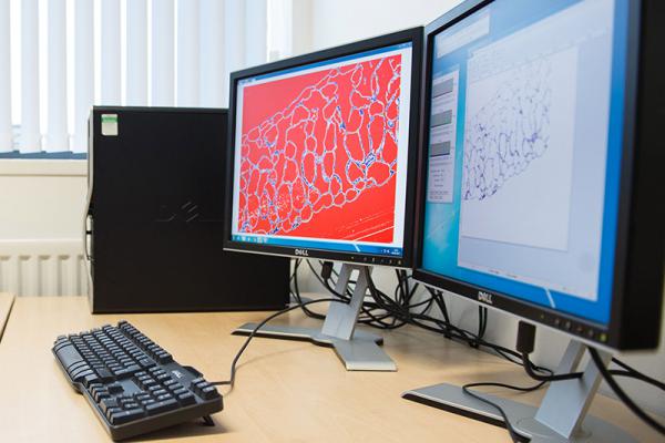
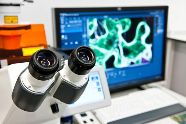
Equipped with three work stations, software including Metamorph, Zeiss Zen 2011, Image J, and Leica LAS-X are available for further image analysis without having to book a microscope.
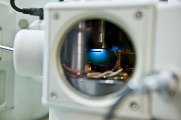
Two of the scanning and transmission electron microscopes are equipped with Oxford EDS x-ray detectors. This allows characterisation of the elemental composition of samples by detecting the x rays emitted by the sample during bombardment by the electron beam. Qualitative and quantitative analysis, and elemental mapping can be performed on both the 6360LV SEM and 2100Plus TEM.
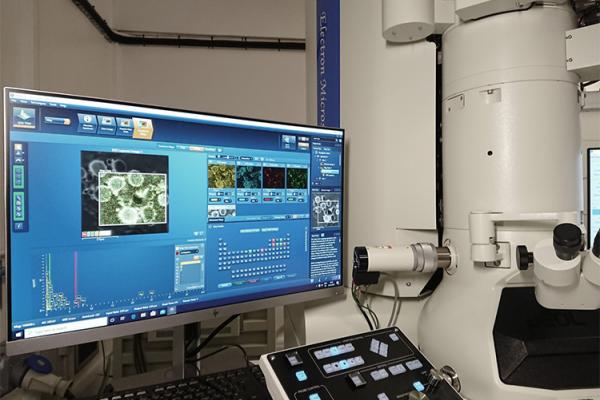
The JEOL 2100Plus transmission electron microscope is a newly commissioned high-resolution TEM capable of reaching 0.25 nm point resolution, and magnifications in excess of 1,200,000x. The TEM is equipped with a Gatan OneView IS camera, capable of acquiring 25 frames per second at full 4k x 4k resolution for high-quality image and video capture. It is fitted with an Oxford Ultim Max 80mm2 EDS detector and dark-field STEM detector to allow high-resolution elemental mapping of nanostructures. High-tilt cryo-tomography holders combined with specialised software packages allow tilted image series to be acquired for the reconstruction of three-dimensional data sets.
The JEOL 6360 scanning electron microscope provides routine imaging of samples at a resolution of 10nm, several hundreds of times greater than optical microscopes. The LV SEM has a Quorum PP3010 cryo-SEM sample preparation system allowing biological specimens to be imaged without dehydration. It also features a low vacuum mode suitable for samples with excessive water content or that cannot be frozen. The backscatter detector provides additional information regarding element composition of the sample.
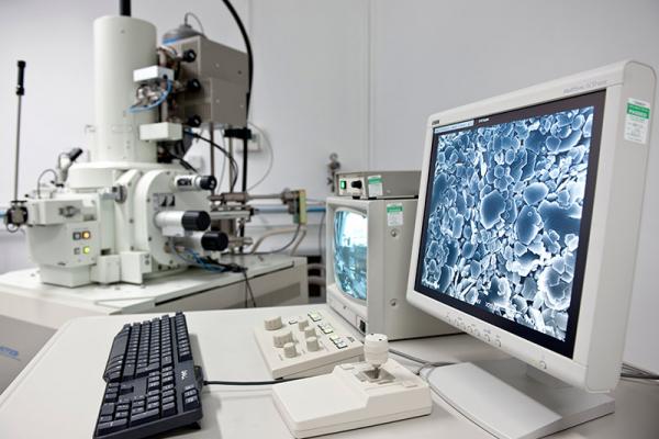
The JEOL 6700F scanning electron microscope uses a cold field emission gun and semi in lens objective to generate high resolution images. It is fitted with a Quorum PP3010 cryo-SEM sample preparation system allowing imaging of biological samples in their native, hydrated state. This SEM has a resolution limit of 3nm and magnification range of 350-100,000x making it suitable for visualising detailed features of specimens.
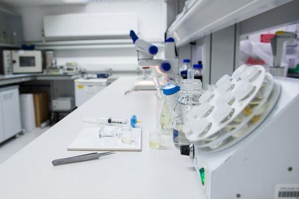
There are two laboratories for sample preparation, allowing users to use Bioimaging for more complex protocols without having to work between two lab spaces. One is used mainly as a clean lab for electron microscopy specimen preparation, the other is well equipped for chemical fixation protocols, wax and resin embedding.
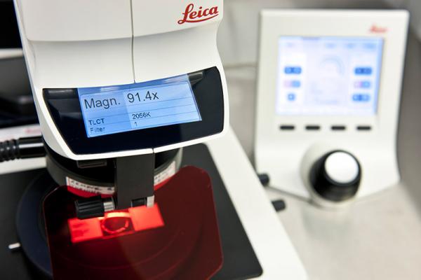
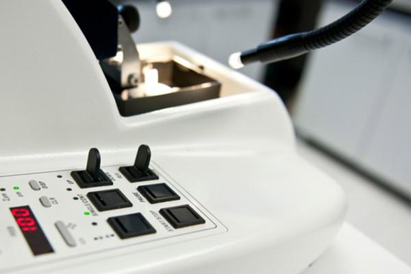
The Leica Cryostat is designed for rapid freezing and sectioning of fresh or fixed tissue samples. The Cryostat will cool from 0 to -35oC and section from 1- 60µm, to give high-quality sections without the need for sample embedding.
The Leica AFS is used to perform freeze substation on high pressure frozen samples. This process is a form of dehydration, removing the water from the sample and replacing it with a solvent prior to resin embedding.
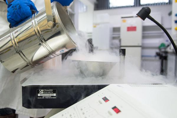
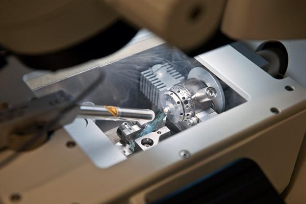
The HPM100 high pressure freezer uses liquid nitrogen and high pressure to rapidly freeze samples as an alternative to chemical fixation that avoids fixation induced artefacts. Samples prepared with the HPM100 are suitable for embedded for both light and electron microscopy.
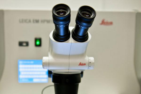
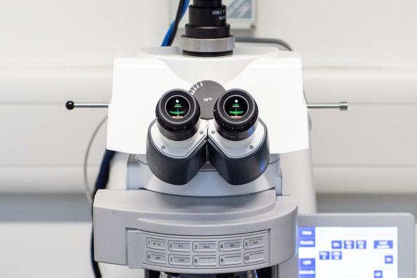
Suitable for observing whole organisms with precise depth of field imaging, the M205 FA is fully motorised to give excellent experiment reproducibility. The M205 has a digital camera capable of colour imaging for both reflected or transmitted brightfield light and monochrome imaging for fluorescence. Fluorescence filters available on the M205 FA stereomicroscope: UV, DAPI, Violet, CFP, GFP, GFP plus, GFP plants, YFP, TxRed, DsRed and mCherry.
Lecia Stellaris 8 Falcon (FAst Lifetime CONtrast) Confocal Laser Microscope with up to 120 nm resolution, has a fully tuneable white light laser ranging from 440nm to 790nm. The system has 5 detectors with either analogue or photon counting modes, allowing imaging into near infra-red wavelengths as well as supporting FLIM, FRET and FRAP imaging techniques.
Thick sections are defined as tissue sections of 0.5µm thickness or above, and are mainly suitable for light and laser confocal microscopy. Samples can be either fresh or fixed and embedded in wax or resin before sectioning.
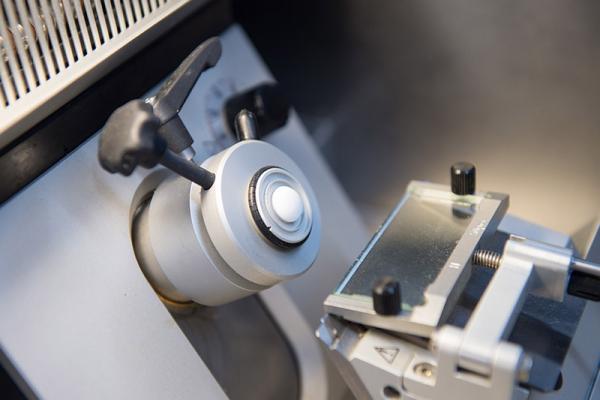
Semi-thin (0.5µm -100nm) and ultra-thin sections (100nm or less) are cut from samples that are resin embedded using a glass or diamond knife. Semi-thin sections can be viewed with a light microscope, but ultra-thin are only suitable for transmission electron microscopy. Cryo-sectioning is a similar technique for thin sectioning samples that have been frozen between -20oC and -185oC, without the requirement of embedding in resin.
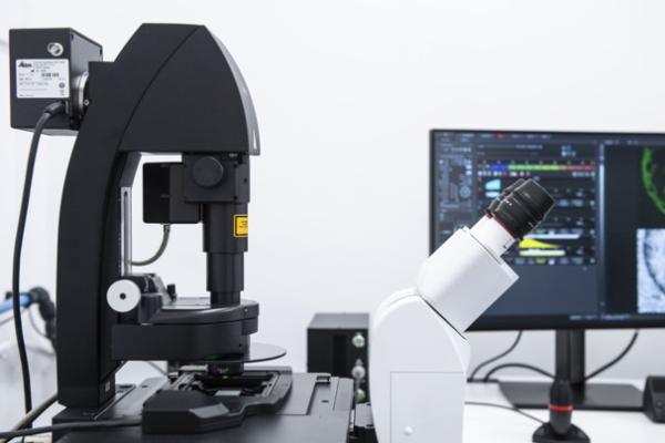
The Axioimager is a fully integrated microscopy system for high resolution light and fluorescence imaging. With a fully motorised XYZ stage and contrast management, high quality images are easily achieved with high reproducibility using the ZEN image software. Zeiss plan-Apochromat objectives with the best colour correction and highest numerical apertures, give high resolution images which can be captured with the 12-megapixel Zeiss Axiocam 512 colour camera. Illumination techniques include brightfield, darkfield, DIC and epifluorescence with filters ranging from DAPI to mRFP.
The Zeiss LSM780 confocal microscope is used for live cell imaging, multi-fluorescence, spectral imaging and unmixing, co-localisation, FRET, FRAP and FLIP. The high sensitivity of LSM 780 is achieved with the 32-channel GaAsP detector which significantly benefits the imaging of plant systems and low signal strength fluorescence.